Muzammal Shafique
MS in Computer Science, University of Michigan, USA · BS in Mathematics from National University of Sciences and Technology, Pakistan
Hi, I am Muzammal Shafique, an MS student in Computer Science at the University of Michigan. I am a Graduate Research Assistant at the SMILES Lab, supervised by Prof. Khalid M. Malik.
Previously, I attained my BS in Mathematics from the National University of Sciences and Technology (NUST), Islamabad, Pakistan.
My broader research interests lies at the intersection of 3D medical image analysis, computer vision, vision foundation models and Phyiscs Informed Learning. I focus on developing data-efficient and generalizable frameworks that leverage VLMs and LLMs for medical imaging, with a particular emphasis on multimodal learning and cerebrovascular and cardiovascular blood hemodynamics prediction.
My active research interests centers on intracranial aneurysm analysis — combining foundation models to enable structure-aware detection, segmentation, and rupture risk prediction across 3D CTA, ToF-MRA, and DSA modalities AND Multimodal Physics-Informed Graph Neural Networks for Intracranial Aneurysm Hemodynamics and Rupture Risk Prediction. View project →
My work has been published at venues including CVPR and ISBI, and has been supported by multiple competitive awards including the University of Michigan Conference Travel Award and the Prime Minister's Ehsaas Undergraduate Scholarship.
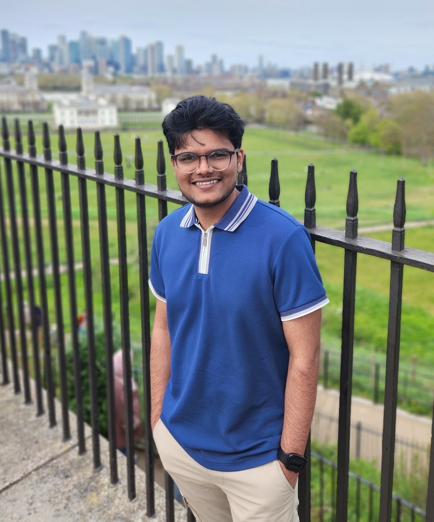
📍Greenwich Maritime, London, UK
Research Interests
Masters Thesis
Towards Reliable Intracranial Aneurysm Analysis: Leveraging Foundation Models for Structure-Aware Detection and Segmentation across 3D CTA, ToF-MRA and DSA
News
| June 2026 | Presenting our paper at CVPR 2026 — awarded a fully sponsored trip including paper registration. 🎉 |
| April 2026 | Awarded University of Michigan Conference Travel Award ($5,000) to attend and present at ISBI 2026 in London, UK. 🎉 |
| March 2026 | Received $1,000 award at the AI in Research Symposium 2026, one of University of Michigan's most prestigious recognition programs. 🎉 |
Selected Publications
-
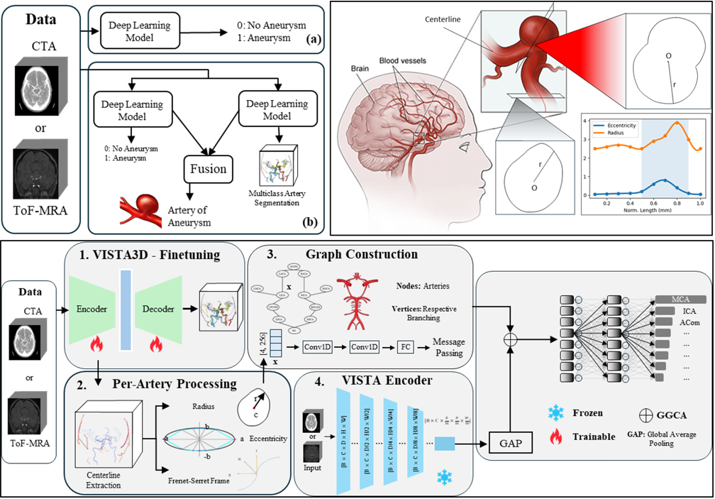 ARAN: Leveraging Foundation Models for Vasculature-Tree-Informed ARtery-Aware Intracranial ANeurysm Detection in 3D CTA and ToF-MRAIn Proceedings of the IEEE/CVF Conference on Computer Vision and Pattern Recognition (CVPR), 2026CVPR 2026Computed Tomography Angiography and Time-of-Flight Magnetic Resonance Angiography serve as clinical standards for non-invasive intracranial aneurysm assessment; however, automated detection and precise localization remain significant challenges. Existing computational methods treat aneurysm detection as a binary detection task. Critically, these approaches typically neglect the identification of the parent artery, a vital parameter for hemodynamic rupture risk assessment and surgical planning. To address these limitations, we present ARAN, an ARtery-aware intracranial ANeurysm detection framework that leverages foundation model representations and vascular geometry priors from CTA and ToF-MRA. Our approach combines a finetuned VISTA3D encoder for volumetric feature extraction with a graph-based geometric branch that models artery specific geometric descriptors, including radius profiles, eccentricity, and Frenet-Serret’s curvature-torsion sequences derived from vessel centerlines. The two branches are fused through Geometry-Gated Cross-Attention, enabling geometry-aware modulation of visual features. Experiments on publicly available datasets demonstrate state of-the-art artery-level classification accuracy of 78.2% on CTA and 85.8% on ToF-MRA, outperforming vision-only and geometry-only baselines.
 -
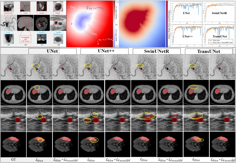 FocusSDF: Boundary-Aware Learning for Medical Image Segmentation via Signed Distance SupervisionIn Proceedings of the 23rd IEEE International Symposium on Biomedical Imaging (ISBI), 2026ISBI 2026 PDFSegmentation of medical images constitutes an essential component of medical image analysis, providing the foundation for precise diagnosis and efficient therapeutic interventions in clinical practices. Despite substantial progress, most segmentation models do not explicitly encode boundary information; as a result, making boundary preservation a persistent challenge in medical image segmentation. To address this challenge, we introduce FocusSDF, a novel loss function based on the signed distance functions (SDFs), which redirects the network to concentrate on boundary regions by adaptively assigning higher weights to pixels closer to the lesion or organ boundary, effectively making it boundary aware. To rigorously validate FocusSDF, we perform extensive evaluations against five state-of-the-art medical image segmentation models, including the foundation model MedSAM, using four distance-based loss functions across diverse datasets covering cerebral aneurysm, stroke, liver, and breast tumor segmentation tasks spanning multiple imaging modalities. The experimental results consistently demonstrate the superior performance of FocusSDF over existing distance transform based loss functions.
 -
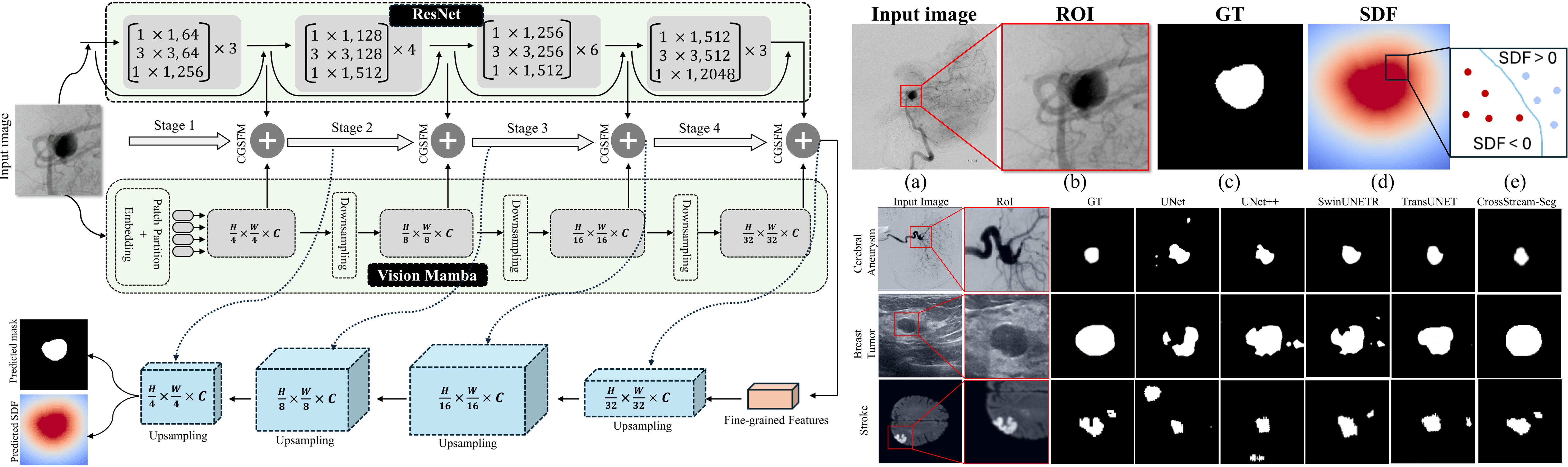 CrossStream-Seg: A Cross-Guided Two-Stream Learning for Boundary-Sensitive ROI SegmentationIn Proceedings of the IEEE/CVF Conference on Computer Vision and Pattern Recognition (CVPR), 2026CVPR 2026Accurate region of interest segmentation in medical images requires both precise local boundary modeling and sufficient global contextual understanding, yet existing architectures often emphasize one at the expense of the other. We propose CrossStream-Seg, a boundary-sensitive ROI encoder built on a cross-guided two-stream encoder module that integrates ResNet-based local representation learning and Vision Mamba-based global contextual modeling. To enable effective interaction between the two streams, we introduce a Cross-Gated Stage Fusion Module, which performs feature alignment, selective cross-stream refinement, and fused multi-scale feature generation throughout the encoder hierarchy. The resulting features are decoded through a shared segmentation pathway and trained with boundary-aware signed-distance supervision to improve geometric consistency near challenging boundaries. The proposed design remains lightweight and structurally simple while explicitly addressing both representation alignment and boundary sensitivity. Experimental results on cerebral aneurysm in DSA, stroke in MRI, and breast tumor in ultrasound imaging modalities show that CrossStream-Seg consistently improves both overlap-based and boundary-aware metrics over counterpart baselines including convolution and transformer-based models. The results indicate that combining structured local-global feature interaction with boundary-aware geometric supervision provides a robust and general framework for medical region of interest segmentation.